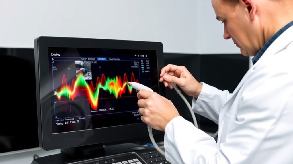
Doppler ultrasounds are performed over 30 million times annually worldwide. This non-invasive imaging technique uses sound waves to measure blood flow and assess conditions related to the heart and blood vessels. You might wonder about safety, preparation, or how it differs from other imaging methods. It’s important to understand these aspects to make informed decisions about your health. Let’s explore the essentials and uncover what this procedure entails.
How Does Doppler Ultrasound Work?
Although the concept may seem complex, Doppler ultrasound technology is remarkably intuitive. You’ll appreciate how it measures blood flow using high-frequency sound waves. The ultrasound transducer sends these waves into your body, which then bounce off moving blood cells.
By analyzing the frequency shift of these echoes, the Doppler system calculates blood flow velocity. This process provides real-time data about the speed and direction of blood flow in your vessels.
Calculating blood flow velocity offers real-time insights into vessel speed and direction.
Understanding this can help you grasp how Doppler ultrasound identifies issues like blockages or abnormal blood flow patterns, which are critical for diagnosing various conditions.
This non-invasive technique offers precise insights without requiring surgical intervention, making it a valuable tool in your healthcare management. Trust in this technology for accurate, timely assessments.
What Are the Different Types of Doppler Ultrasound?
Having understood how Doppler ultrasound operates, it’s important to explore the various types available, each suited to specific diagnostic needs.
Color Doppler converts sound waves into colors to visualize blood flow direction and speed, providing a thorough overview.
Power Doppler, though similar, offers more sensitivity for detecting low-velocity blood flow, especially beneficial in small vessels.
Spectral Doppler, on the other hand, focuses on providing detailed data about blood flow velocity through graphs, aiding in precise assessments.
Continuous wave Doppler excels in measuring high-speed blood flow, essential for evaluating conditions like heart valve issues.
Meanwhile, pulsed wave Doppler provides accurate flow measurements at specific locations, enhancing the diagnostic capability.
What Conditions Can Be Diagnosed With Doppler Ultrasound?
When it comes to diagnosing medical conditions, Doppler ultrasound is an invaluable tool in your healthcare arsenal. It uses sound waves to evaluate blood flow through your vessels, identifying issues like blockages or narrowed arteries. This non-invasive procedure helps diagnose various conditions with precision and reliability.
| Condition | Diagnostic Insight |
|---|---|
| Peripheral Artery Disease | Detects reduced blood flow in your extremities. |
| Deep Vein Thrombosis (DVT) | Identifies blood clots in your deep veins. |
| Carotid Artery Stenosis | Assesses narrowing in your neck arteries, preventing stroke. |
| Venous Insufficiency | Evaluates improper blood flow in your veins. |
Using Doppler ultrasound, you can address these conditions early, reducing potential complications. Whether you’re experiencing symptoms or undergoing routine checks, this technology plays a vital role in maintaining your vascular health.
How Is Doppler Ultrasound Used in Pregnancy?
During pregnancy, you can rely on Doppler ultrasound to effectively monitor fetal blood flow, ensuring the baby receives adequate oxygen and nutrients.
This technology also assesses placental health by measuring blood flow in the uterine and umbilical arteries, which helps identify potential complications.
Additionally, Doppler ultrasound plays an essential role in evaluating fetal growth, allowing your healthcare provider to make informed decisions about your pregnancy care.
Monitoring Fetal Blood Flow
How exactly does Doppler ultrasound assist in monitoring fetal blood flow during pregnancy? It measures blood velocity in fetal vessels, providing essential data about circulatory health. You’ll find this non-invasive tool invaluable in evaluating conditions like intrauterine growth restriction (IUGR) or fetal distress. By capturing real-time images, Doppler ultrasound helps you guarantee your baby’s well-being by examining blood flow in the umbilical artery, middle cerebral artery, and ductus venosus.
| Parameter | Purpose |
|---|---|
| Umbilical Artery | Evaluates placental and fetal circulation |
| Middle Cerebral Artery | Examines fetal brain blood flow |
| Ductus Venosus | Monitors venous blood return |
| Uterine Artery | Checks maternal-fetal blood exchange |
| Cardiac Output | Observes fetal heart function |
Incorporating these analyses lets you proactively manage your pregnancy, guaranteeing ideal fetal health.
Assessing Placental Health
Although Doppler ultrasound is widely used for various purposes in pregnancy, its role in evaluating placental health is particularly significant. You’ll find it helps assess blood flow in the umbilical and uterine arteries, offering vital insights into placental function.
By measuring resistance indices, it identifies abnormalities such as placental insufficiency, which can impact fetal well-being. This non-invasive technique allows healthcare providers to monitor conditions like preeclampsia or placental abruption, ensuring timely interventions.
When you undergo a Doppler ultrasound, the sonographer evaluates the waveforms of blood flow, which provide data on how well the placenta is supplying nutrients and oxygen.
Understanding these patterns helps your care team make informed decisions, tailoring your prenatal care and potentially improving pregnancy outcomes.
Evaluating Fetal Growth
Doppler ultrasound plays an essential role in evaluating fetal growth by measuring blood flow in key fetal vessels such as the umbilical artery and middle cerebral artery.
These measurements provide vital insights into your baby’s well-being. By examining the resistance in these vessels, Doppler ultrasound helps determine if your baby is receiving adequate oxygen and nutrients.
This technique is especially important if there’s concern about intrauterine growth restriction (IUGR) or other complications. When abnormalities in blood flow are detected, your healthcare provider can develop targeted interventions to support ideal fetal development.
Regular Doppler evaluations can guide decisions about the timing of delivery and necessary treatments, ensuring you and your baby get the best possible care throughout your pregnancy.
What Should Patients Expect During the Procedure?
Before your Doppler ultrasound, you may be asked to change into a gown and remove any jewelry that could interfere with the procedure.
During the ultrasound, you’ll lie on an examination table while a technician applies a conductive gel to your skin to guarantee clear transmission of sound waves.
The technician will then use a transducer to capture real-time images and assess blood flow, providing valuable diagnostic information.
Procedure Preparation Steps
Preparing for a Doppler ultrasound involves several straightforward steps to guarantee accurate results.
First, follow any specific instructions your healthcare provider gives, as they may vary depending on the area being examined. You might need to fast or avoid consuming caffeine, which can affect blood flow readings.
Wear loose, comfortable clothing, as you may need to change into a gown. Remove any jewelry or metallic objects that could interfere with the ultrasound waves.
Arrive at the facility a few minutes early to complete any necessary paperwork and relax before the procedure. Keep a list of current medications handy, as the technician might ask about them.
If you’re on blood thinners, inform the medical team, as these can impact the ultrasound’s effectiveness.
During the Ultrasound Experience
Once you’ve completed the preparation steps, you’ll be ready for the Doppler ultrasound procedure itself.
During the examination, you’ll lie comfortably on an examination table. A trained sonographer will apply a water-based gel to your skin, guaranteeing ideal sound wave transmission.
They’ll then glide a transducer over the targeted area. This handheld device sends and receives high-frequency sound waves, creating real-time images of blood flow within your vessels.
You’ll hear whooshing sounds, indicating blood movement, which is perfectly normal. The procedure is non-invasive and painless, though you may feel slight pressure from the transducer.
Stay relaxed and still to guarantee precise results. Typically, the process lasts 30 to 60 minutes, after which you can resume regular activities without restrictions.
Are There Any Risks or Side Effects?
Doppler ultrasound is generally considered safe and non-invasive, making it a preferred diagnostic tool for many medical professionals. It uses high-frequency sound waves to evaluate blood flow through your vessels, avoiding ionizing radiation.
As a patient, you’ll be relieved to know there are no known risks or side effects associated with this technique. It’s painless and usually doesn’t require any special precautions.
During the procedure, a trained sonographer will apply a gel to your skin to improve sound wave transmission. You might feel slight pressure as the transducer moves over your body, but it’s typically comfortable.
Rest assured, the equipment adheres to strict safety standards. If you have any specific concerns, discuss them with your healthcare provider for personalized advice.
How to Prepare for a Doppler Ultrasound Examination?
Before undergoing a Doppler ultrasound examination, it’s essential to follow specific preparation guidelines to guarantee accurate results.
Begin by wearing comfortable, loose-fitting clothing; it simplifies access to the area being examined. Depending on the test location, you might need to avoid eating or drinking several hours before the procedure. Your healthcare provider will inform you of any dietary restrictions.
Wear comfortable clothing and follow dietary restrictions if necessary for accurate Doppler ultrasound results.
If you’re on medication, continue taking it unless instructed otherwise. It’s important to disclose any known allergies, particularly to latex, as the technician might use a gel during the exam.
Arrive early to complete any necessary paperwork, and bring a list of medications and medical history. Following these steps guarantees a smooth, efficient Doppler ultrasound process with maximum diagnostic accuracy.
How Does Doppler Ultrasound Compare to Other Imaging Techniques?
As you prepare for a Doppler ultrasound, understanding how it stacks up against other imaging techniques can enhance your appreciation of its unique benefits.
Unlike CT or MRI, Doppler ultrasound uses sound waves instead of radiation, ensuring a safer option for examining blood flow and vascular conditions. It offers real-time imaging, capturing dynamic blood movement, which is vital for diagnosing blockages or clots.
While MRI provides detailed soft tissue visualization, Doppler excels in evaluating hemodynamics without contrast agents. Compared to X-rays, it’s non-invasive and doesn’t require ionizing radiation.
Although it may not visualize deep tissue structures as effectively as an MRI, its portability and cost-effectiveness make it ideal for bedside evaluations and monitoring vascular health efficiently.
Frequently Asked Questions
How Long Does a Doppler Ultrasound Examination Typically Take?
A Doppler ultrasound examination typically takes 30 to 60 minutes. You’ll lie comfortably while a technician uses a transducer to assess blood flow. It’s non-invasive, so you’ll experience minimal discomfort, and results are usually available quickly.
Is a Doppler Ultrasound Examination Covered by Insurance?
Yes, most insurance plans cover Doppler ultrasound exams, considering them medically necessary. You should confirm coverage with your provider, ensuring your specific policy includes this diagnostic procedure. Always check for any copayments or deductibles applicable.
Can Doppler Ultrasound Detect Deep Vein Thrombosis (DVT)?
Yes, a Doppler ultrasound effectively detects deep vein thrombosis by using sound waves to visualize blood flow. You’ll appreciate its non-invasive nature, providing real-time, accurate insights into potential blood clots, ensuring timely, appropriate management.
Are There Any Dietary Restrictions Before a Doppler Ultrasound?
No, you don’t need to follow any dietary restrictions before a Doppler ultrasound. The procedure is non-invasive and doesn’t require special preparation. Just wear comfortable clothing to guarantee easy access to the area being examined.
Can Medications Affect the Results of a Doppler Ultrasound?
Yes, medications can influence Doppler ultrasound results. Like a conductor guiding an orchestra, certain drugs may alter blood flow, affecting the imaging. Discuss your current medications with your healthcare provider before the procedure for accurate results.
Conclusion
Doppler ultrasound offers a safe, non-invasive way to assess blood flow, and you’ll find it generally painless, experiencing only mild pressure. It’s versatile in diagnosing conditions, from vascular issues to monitoring fetal health. Notably, it’s used in about 70% of pregnancies to check fetal well-being. Before your exam, consult your healthcare provider about any dietary or medication adjustments. Compared to other imaging techniques, Doppler ultrasound provides real-time insights without radiation exposure, ensuring peace of mind.
