When performing a Doppler ultrasound, you’re responsible for guaranteeing accurate and reliable results by adhering to specific do’s and don’ts. Position your patient correctly and communicate clearly to enhance their comfort and cooperation. Calibrate your equipment meticulously and verify settings to maintain precision. Avoid excessive pressure and incorrect angles to prevent skewed readings. As you continue, you’ll explore more nuances that will refine your skills and guarantee patient trust and safety.
Understanding Doppler Ultrasound Basics
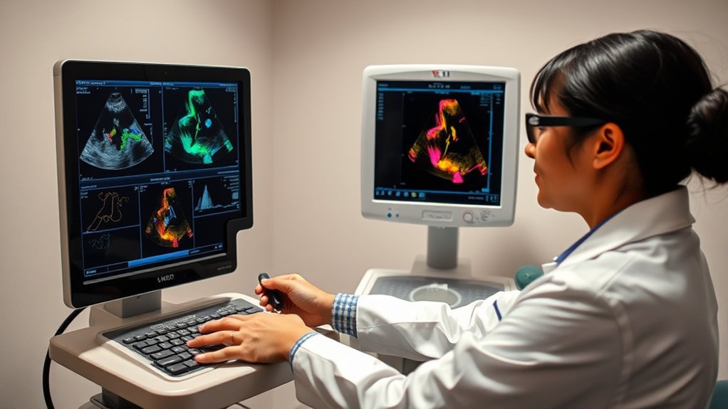
Doppler ultrasound technology leverages sound waves to measure and visualize blood flow within vessels, offering invaluable insights into cardiovascular health. You need to understand how it works. The device emits high-frequency sound waves that bounce off circulating red blood cells. The reflected waves are detected and used to create images and graphs, illustrating the speed and direction of blood flow.
You should know that Doppler ultrasound identifies abnormalities like blockages, clots, or narrowed arteries, which may indicate conditions like deep vein thrombosis or carotid artery stenosis.
Engage with the data critically; observe the flow patterns and velocities. In addition, guarantee ideal probe placement for accurate readings. Understanding these basics equips you to recognize potential cardiovascular issues early and take appropriate action.
Preparing for a Doppler Ultrasound Exam
Before you undergo a Doppler ultrasound exam, make sure you’re adequately prepared to maximize the test’s accuracy.
Start by consulting your physician for any specific instructions related to your medical condition, as preparation may vary. It’s essential to adhere to these guidelines to guarantee precise results:
- Hydration: Drink plenty of water unless instructed otherwise, as it aids in clearer imaging.
- Medication: Continue taking prescribed medications unless your doctor advises a temporary pause.
- Fasting: If instructed, avoid eating or drinking (besides water) for a specified period prior to the exam.
- Clothing: Wear loose, comfortable clothing to facilitate easy access to the area being examined.
Following these steps will help optimize your Doppler ultrasound experience.
Best Practices for Accurate Results
To guarantee accurate results during a Doppler ultrasound, start by positioning the patient correctly to optimize vascular access and minimize artifacts.
Calibrate the equipment meticulously, verifying settings like frequency and gain to suit the specific examination.
Minimize patient movement by providing clear instructions and assuring comfort, as motion can lead to inaccurate measurements.
Proper Patient Positioning
For ideal Doppler ultrasound results, ensuring proper patient positioning is essential. Start by adjusting the examination table to the correct height, allowing you to maintain ergonomic posture while scanning.
Positioning not only affects your comfort but also the accuracy of the readings. Here’s what you should do:
- Supine Position: For abdominal and lower extremity exams, have the patient lie flat with a slight head elevation.
- Left Lateral Decubitus: Use this for cardiac assessments; it helps optimize heart visualization.
- Sitting Position: Effective for carotid artery scans by extending the neck slightly.
- Prone Position: Beneficial for scanning the posterior aspect, such as certain vascular exams.
Ensure the patient is comfortable, and always communicate the positioning process clearly to enhance cooperation and relaxation.
Equipment Calibration Essentials
Proper patient positioning lays the groundwork for successful Doppler ultrasound examinations, but even the best positioning can’t compensate for inaccurate equipment.
Begin by checking your device’s calibration regularly. Verify frequency settings match manufacturer specifications. Confirm that the Doppler angle aligns correctly; typically, an angle below 60 degrees yields ideal results.
Adjust the gain settings to verify signals aren’t excessively amplified or diminished. Always verify the transducer’s integrity—clean and inspect it for any wear.
Conduct a test run on a known phantom model to confirm accuracy before patient use. Document calibration results meticulously. Keep software up-to-date for enhancements and error corrections.
Handling Patient Movement
When conducting a Doppler ultrasound, managing patient movement is essential to obtaining accurate results. You need to guarantee the patient remains as still as possible during the procedure. To achieve this, follow these best practices:
- Communicate Clearly: Explain the procedure and the importance of remaining still. This helps set patient expectations and reduces anxiety.
- Position Comfortably: Adjust the patient’s position to ensure comfort and stability, minimizing involuntary movements.
- Use Supports: Utilize pillows or supports to stabilize limbs or other body parts that may shift during the examination.
- Monitor Continuously: Keep a watchful eye on the patient throughout the procedure, addressing any discomfort or adjustments needed promptly.
With these techniques, you can greatly enhance the reliability of your Doppler ultrasound results.
Common Mistakes to Avoid
In the domain of Doppler ultrasound, technicians often encounter several common mistakes that can compromise the accuracy of diagnostic results.
First, guarantee ideal transducer positioning. Misalignment can lead to inaccurate velocity readings. Always align the transducer parallel to blood flow. Avoid excessive pressure, as it may distort blood vessels, affecting measurements.
In addition, improper gain settings can obscure or exaggerate signals. Adjust gain to eliminate noise while preserving signal integrity.
Don’t forget to verify the angle of insonation; angles greater than 60 degrees can lead to significant errors in velocity estimation.
Finally, neglecting to calibrate equipment can result in systemic errors. Regularly check and calibrate your machine to maintain reliability.
Patient Communication and Comfort
Beyond technical accuracy, patient comfort and effective communication are essential in Doppler ultrasound procedures. You should guarantee that patients are at ease and well-informed throughout the process.
Ensure patient comfort and clear communication during Doppler ultrasound for a seamless and reassuring experience.
Begin by providing a clear explanation of the procedure, emphasizing key points without overwhelming them.
- Maintain Eye Contact: This builds trust and assures the patient you’re attentive to their needs.
- Use Simple Language: Avoid medical jargon; keep explanations straightforward and understandable.
- Invite Questions: Encourage patients to ask questions, guaranteeing they feel heard and understood.
- Comfortable Positioning: Adjust the patient’s position for maximum comfort, minimizing any potential discomfort during the procedure.
Interpreting Doppler Ultrasound Findings
How do you certify accurate interpretation of Doppler ultrasound findings?
First, make sure you’re familiar with normal hemodynamic patterns. Recognize the spectral waveform characteristics, such as peak systolic velocity and end-diastolic velocity. Identify any deviations from expected patterns, noting turbulence or aliasing, which might indicate pathological changes.
Always cross-reference findings with patient history and clinical data for context. Use standardized protocols to measure and report velocities consistently. Remember, angle correction is essential for accurate velocity estimations—keep the Doppler angle as close to 60 degrees as possible.
Evaluate both qualitative and quantitative data, and compare with established reference values. Collaborate with colleagues for complex cases and continually update your knowledge through ongoing education and training.
This structured approach guarantees reliability in your interpretations.
Ensuring Safety and Minimizing Risks
Accurate interpretation of Doppler ultrasound findings lays the groundwork for effective clinical practice, but guaranteeing patient safety and minimizing risks is paramount.
Prioritize these practices to safeguard patients and enhance diagnostic efficacy:
- Use the lowest possible power settings to achieve the diagnostic objectives, reducing the risk of thermal and mechanical effects.
- Adhere to ALARA principles (As Low As Reasonably Achievable) for exposure time and intensity to minimize unnecessary exposure.
- Regularly calibrate and maintain equipment to guarantee peak performance and accurate readings, preventing potential diagnostic errors.
- Educate patients on the procedure, addressing any concerns to foster cooperation and ease anxiety, thereby guaranteeing smoother examinations.
Frequently Asked Questions
Can Doppler Ultrasound Detect Cancer?
No, Doppler ultrasound can’t directly detect cancer. It evaluates blood flow patterns, helping identify abnormal vascular activity often associated with tumors. Use it in conjunction with other diagnostic tools for a thorough assessment of potential malignancies.
Is Doppler Ultrasound Safe During Pregnancy?
Yes, Doppler ultrasound is safe during pregnancy, as it uses sound waves, not radiation. You might worry about effects, but studies show no adverse risks. Always consult your healthcare provider for personalized guidance and safe practices.
How Does Doppler Ultrasound Differ From Traditional Ultrasound?
Doppler ultrasound measures blood flow velocity, while traditional ultrasound provides structural imaging. You’ll use Doppler to assess circulation in vessels or organs, whereas traditional ultrasound captures static images for anatomical analysis. Remember, adjust settings for best results.
Can Doppler Ultrasound Assess Heart Valve Function?
Yes, you can use Doppler ultrasound to assess heart valve function. While traditional ultrasounds show structure, Doppler reveals blood flow. Evaluate velocity and direction to detect abnormalities like stenosis or regurgitation. It’s precise, efficient, diagnostic.
Are There Any Dietary Restrictions Before a Doppler Ultrasound?
You don’t need to follow any specific dietary restrictions before a Doppler ultrasound. However, for abdominal scans, it’s best to fast for 6-8 hours. Always consult your healthcare provider for personalized preparation instructions beforehand.
Conclusion
In mastering Doppler ultrasound, remember: precision is your ally and patient comfort, your guide. Calibrate equipment meticulously, position patients thoughtfully, and communicate clearly—these actions forge the path to accurate results. Avoid pitfalls like excessive vessel pressure and improper insonation angles, which can distort findings. By fostering an environment of understanding and trust, you not only enhance the exam’s effectiveness but also alleviate patient anxiety. With these practices, you’re not just performing a test; you’re crafting a diagnostic masterpiece.