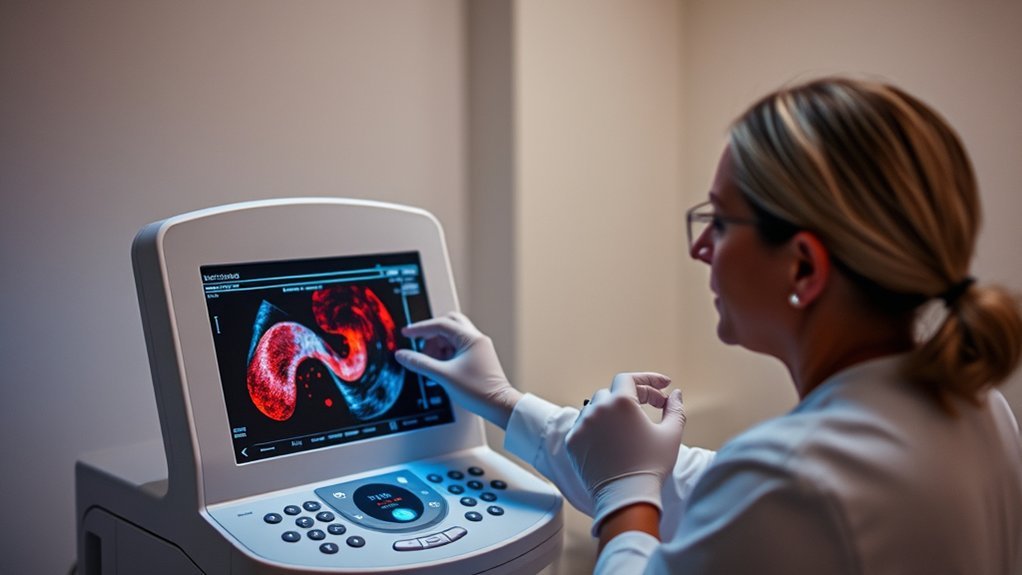
You might think Doppler ultrasound is just for pregnancy or worry about radiation risks, but these are misconceptions. This non-invasive technique uses sound waves for evaluating blood flow, proving essential in various medical fields. It’s safe, painless, and has evolved with technology, enhancing its diagnostic capabilities. Understanding its accuracy and safety could change your perception. Curious to learn how it actually functions and its true applications?
Understanding Doppler Ultrasound and Its Functionality
When you explore the world of Doppler ultrasound, it’s essential to grasp its core functionality and how it differs from traditional ultrasound.
Doppler ultrasound employs the Doppler effect to measure and visualize blood flow dynamics within vessels. Unlike traditional ultrasound, which primarily provides static structural images, Doppler ultrasound detects shifts in frequency caused by the movement of red blood cells.
This capability allows you to assess the velocity and direction of blood flow, aiding in the diagnosis of vascular conditions.
Debunking the Myth: Doppler Ultrasound and Radiation Exposure
Having established the fundamental workings of Doppler ultrasound, it’s important to address a common misconception: the belief that Doppler ultrasound exposes patients to radiation. This isn’t accurate.
Doppler ultrasound employs sound waves, not ionizing radiation like X-rays or CT scans. The ultrasound transducer emits high-frequency sound waves that bounce off moving blood cells. These sound waves are then converted into real-time images, providing valuable insights into blood flow.
Doppler ultrasound uses sound waves, not radiation, for real-time blood flow imaging.
Scientific evidence confirms that Doppler ultrasound is a safe diagnostic tool, free from radiation risks. It’s essential to understand that the technology hinges on acoustics, not radiation physics.
Therefore, when considering Doppler ultrasound for medical evaluations, you can rest assured it doesn’t involve any radiation exposure, ensuring patient safety and peace of mind.
The Role of Doppler Ultrasound in Diagnosing Blood Flow Issues
In diagnosing blood flow issues, Doppler ultrasound serves as an invaluable tool, offering detailed insights into the circulatory system’s functionality. You’ll find it essential for evaluating vascular health, detecting abnormalities, and guiding treatment plans. This non-invasive method measures blood flow velocity with precision, identifying conditions like arterial blockages and venous insufficiencies.
| Doppler Type | Key Application | Benefit |
|---|---|---|
| Continuous Wave | High-speed blood flow | Identifies stenosis |
| Pulsed Wave | Specific location focus | Localized flow evaluation |
| Color Doppler | Visual flow representation | Quick anomaly detection |
| Power Doppler | Sensitive flow detection | Detects low-velocity flows |
Exploring the Accuracy of Doppler Ultrasound Results
To accurately assess Doppler ultrasound results, you need a solid grasp of Doppler mechanics and their role in interpreting blood flow data.
Factors such as transducer frequency and angle can influence result precision, so it’s essential to account for these variables.
Understanding Doppler Mechanics
A key aspect of understanding Doppler ultrasound mechanics lies in its ability to measure the velocity of blood flow with remarkable precision. You can achieve this through the Doppler effect, which detects frequency shifts when ultrasound waves bounce off moving red blood cells. By analyzing these shifts, you determine blood flow velocity and direction.
This non-invasive method relies on advanced algorithms and high-frequency sound waves to provide real-time, accurate data.
Doppler ultrasound’s precision is influenced by factors like the angle between the ultrasound beam and blood flow. Ideally, you should maintain an angle of 60 degrees or less to optimize accuracy.
Additionally, modern Doppler devices incorporate technologies like color flow mapping, enhancing your ability to visualize and assess hemodynamic conditions efficiently.
Interpreting Ultrasound Results
While examining Doppler ultrasound results, it’s crucial to grasp the nuances of accuracy and interpretation. Doppler ultrasound effectively evaluates blood flow dynamics, providing critical insights into vascular health.
You need to accurately interpret spectral waveforms and color flow images to assess blood velocity and direction. Recognize that accuracy can vary based on the operator’s skill and equipment calibration. Always correlate findings with clinical context and other diagnostic tests.
For precise results, pay attention to angle correction, as errors can skew velocity measurements. Make sure the Doppler angle is less than 60 degrees for peak accuracy.
Familiarize yourself with normal and pathological waveforms to identify abnormalities effectively. Staying current with guidelines and advances in Doppler technology will enhance your ability to provide accurate interpretations.
Accuracy Influencing Factors
Understanding the nuances of interpreting Doppler ultrasound results naturally leads to exploring the factors influencing their accuracy. Several essential elements can considerably impact the reliability of these results.
First, operator expertise plays a fundamental role. Skilled professionals are more likely to produce precise readings.
Second, equipment quality can’t be underestimated. High-quality machines yield more reliable data.
Third, patient factors, such as movement or tissue characteristics, can introduce variability.
Finally, environmental conditions, like room temperature or ambient noise, may subtly affect outcomes.
Consider the emotional consequences if:
- Operator error causes misdiagnosis.
- Outdated equipment leads to inaccurate results.
- Patient discomfort affects the scanning process.
- External distractions compromise the exam’s integrity.
Safety Considerations for Patients Undergoing Doppler Ultrasound
Even though Doppler ultrasound is widely regarded as a safe diagnostic tool, it’s important to evaluate specific safety aspects for patients.
As a non-invasive procedure, it utilizes sound waves rather than ionizing radiation, minimizing potential risks. You’re unlikely to experience any discomfort, as the process doesn’t require penetration or incisions.
However, it’s essential to ascertain the equipment is properly calibrated, as incorrect settings may lead to inaccurate results or unnecessary exposure to energy. When performed by trained professionals, the risk of thermal or mechanical effects is negligible.
Always inform your healthcare provider of any implantable devices, as they may influence the procedure.
Ultimately, Doppler ultrasound’s safety profile is robust, providing critical diagnostic information without significant adverse effects.
Common Misconceptions About Doppler Ultrasound in Pregnancy
Misconceptions surrounding Doppler ultrasound in pregnancy can lead to unnecessary anxiety or misinformation. One prevalent myth is that Doppler ultrasound emits harmful radiation, posing risks to the developing fetus. In reality, Doppler ultrasound uses sound waves, not radiation, ensuring safety when performed correctly.
Doppler ultrasound uses sound waves, not radiation, ensuring fetal safety when used correctly.
Another misunderstanding is that it can replace routine prenatal checkups. While informative, it complements but doesn’t substitute extensive care. Some believe its frequent use may overheat fetal tissues. However, studies show that when used appropriately, it doesn’t cause thermal damage.
Finally, there’s a misconception that it always detects fetal abnormalities. While it aids in evaluating blood flow, it’s not a definitive diagnostic tool for all conditions.
- Fear of harmful radiation
- Misplaced trust as a standalone tool
- Concerns about fetal overheating
- Overestimation of diagnostic capabilities
Frequently Asked Questions
Can Doppler Ultrasound Detect Tumors or Cancerous Growths?
Doppler ultrasound can’t directly detect tumors or cancerous growths. It assesses blood flow, helping identify abnormal vascular patterns often associated with tumors. For a definitive diagnosis, you’d need additional imaging like MRI, CT, or a biopsy.
How Does Doppler Ultrasound Differ From a Standard Ultrasound?
Doppler ultrasound differs by measuring blood flow velocity and direction, using the Doppler effect, whereas standard ultrasound creates structural images. It’s excellent for evaluating vascular conditions, whereas standard ultrasound focuses on anatomy without examining flow dynamics.
Is It Possible to Use Doppler Ultrasound for Heart Condition Monitoring?
Yes, you can use Doppler ultrasound for heart condition monitoring. It evaluates blood flow, detecting abnormalities like valve issues or blockages. This non-invasive method provides real-time data, aiding in accurate diagnosis and effective management of cardiac conditions.
What Are the Cost Considerations for Doppler Ultrasound Procedures?
You’re considering Doppler ultrasound costs, which can vary like night and day. Factors include location, healthcare provider, insurance coverage, and procedure complexity. Typically, costs range from $200 to $1,000. Check with your provider for precise estimates.
Are There Any Alternative Imaging Techniques to Doppler Ultrasound?
Yes, you can consider alternatives like MRI, CT angiography, or traditional ultrasound. Each technique offers unique benefits; MRI provides detailed soft tissue images, CT is fast and thorough, and ultrasound offers convenience and safety.
Conclusion
Think of Doppler ultrasound as a lighthouse guiding ships safely through dark waters. It doesn’t emit harmful radiation; instead, it uses sound waves to illuminate the path of blood flow, highlighting potential obstacles like arterial blockages. Just as a lighthouse aids all vessels, Doppler ultrasound serves diverse medical needs beyond pregnancy. With precision and safety, it reveals hidden currents within the body, ensuring navigational accuracy in diagnosing vascular health, dispelling myths, and fostering informed healthcare decisions.
