Imagine the Doppler ultrasound as a symphony, where each sound wave plays an essential role in revealing the story of blood flow. You need to master the selection of the right transducer and guarantee the patient is correctly positioned for ideal results. Apply coupling gel, adjust settings, and maintain the correct angle. Capture peak systolic and end-diastolic velocities to assess vascular health. Ready to interpret findings and uncover hidden vascular secrets?
Understanding the Basics of Doppler Ultrasound
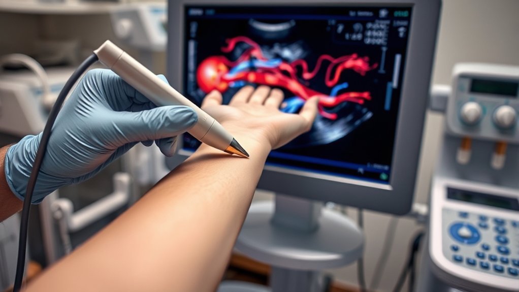
Doppler ultrasound, a powerful diagnostic tool, allows you to visualize and assess blood flow within vessels, utilizing sound waves to measure changes in frequency caused by the movement of red blood cells.
When approaching Doppler ultrasound, first verify the transducer is appropriately selected for the target vessel’s depth and size. Proper patient positioning maximizes vessel exposure and enhances scan accuracy.
You must apply sufficient gel to minimize air gaps between the transducer and the skin, guaranteeing ideal sound wave transmission. Adjust the machine settings, such as gain and scale, to enhance image clarity and flow detection.
Understanding the angle of insonation is vital—maintain it below 60 degrees to avoid significant errors in velocity measurement.
Follow these steps to achieve precise diagnostic results.
Types of Doppler Ultrasound Techniques
Various techniques exist within the domain of Doppler ultrasound, each tailored for specific diagnostic purposes.
You’ll encounter Continuous Wave (CW) Doppler, which measures high-velocity blood flow without aliasing, ideal for cardiac assessments.
Pulsed Wave (PW) Doppler, in contrast, allows you to measure flow at specific depths, providing precise localization but with risk of aliasing in high velocities.
Color Doppler visualizes flow direction and velocity within a region by superimposing color-coded data on the grayscale image, aiding in vascular assessments.
Power Doppler, unlike color Doppler, doesn’t show direction but is more sensitive to low flow states, useful in detecting small vessels.
Tissue Doppler Imaging (TDI) measures myocardial motion, essential for cardiac function evaluation.
Select the technique based on clinical objectives and patient needs.
Equipment and Setup for Doppler Ultrasound
When setting up equipment for a Doppler ultrasound, make certain you have the essential components: a high-quality ultrasound machine with Doppler capabilities, a suitable transducer, and a coupling gel.
Begin by ensuring the ultrasound machine is calibrated for ideal Doppler performance. Select a transducer with the appropriate frequency for the specific vascular assessment—higher frequencies for superficial structures and lower for deeper vessels.
Apply the coupling gel liberally to eliminate air gaps between the transducer and the skin, enhancing sound wave transmission. Position the machine for easy access to controls, ensuring the monitor is visible and the patient area is accessible.
Adjust the machine’s settings, including gain and scale, to enhance image clarity and Doppler signal sensitivity before proceeding.
Preparing the Patient for a Doppler Ultrasound
To guarantee accurate Doppler ultrasound results, you’ll need to instruct the patient on appropriate clothing, ideally loose-fitting garments that allow easy access to the examination area.
Review the patient’s medical history thoroughly to identify any factors that could affect the procedure or results.
Provide clear pre-procedure instructions, including guidelines on fasting or medication adjustments if necessary.
Patient Clothing Requirements
Before undergoing a Doppler ultrasound, it’s essential to confirm the patient is appropriately dressed to facilitate the procedure.
You should instruct the patient to wear loose-fitting, comfortable clothing that allows easy access to the area being examined. Avoid garments with zippers, buttons, or heavy embellishments, as these can interfere with ultrasound waves.
If abdominal or lower extremity scanning is required, suggest wearing a two-piece outfit to allow for easy exposure without complete disrobing.
Instruct the patient to remove any jewelry or accessories near the examination area to prevent artifacts.
For added convenience, provide a gown or drape to maintain patient modesty and comfort during the procedure.
Make sure the patient understands these guidelines to confirm an efficient and accurate Doppler ultrasound examination.
Medical History Review
Besides appropriate attire, a thorough medical history review is necessary to prepare the patient for a Doppler ultrasound.
Start by gathering information on any pre-existing cardiovascular conditions, as these can influence the procedure’s focus. Note any recent surgeries, especially those involving the vascular system, as they can alter blood flow patterns.
It’s vital to document medication usage, including anticoagulants and vasodilators, which might affect vascular imaging. Inquire about symptoms like swelling, pain, or numbness that could indicate underlying vascular issues.
Assess any family history of vascular diseases to identify hereditary risk factors. Confirm if the patient has allergies, especially to ultrasound gel or latex.
This detailed review guarantees you tailor the Doppler ultrasound to the patient’s specific medical needs.
Pre-Procedure Instructions
Although preparing for a Doppler ultrasound might seem straightforward, adhering to specific pre-procedure instructions guarantees the best imaging results.
First, wear loose, comfortable clothing to allow easy access to the area being examined. You may need to remove jewelry or other metal objects, as they can interfere with the ultrasound waves.
Fasting for 6-8 hours prior is vital if the abdominal area is involved, as food can obscure clear imaging. Hydration is important, so drink water unless directed otherwise.
If you’re on medications, consult your healthcare provider, as some might affect blood flow and imaging accuracy.
Arrive at least 15 minutes early to complete any necessary paperwork and guarantee you’re relaxed before the procedure. Your cooperation enhances diagnostic accuracy.
Performing a Doppler Ultrasound Examination
To perform a Doppler ultrasound examination efficiently, verify your equipment is calibrated and settings are optimized for the specific vascular region being assessed.
Utilize precise image acquisition techniques by adjusting the transducer angle and gain settings to enhance blood flow visualization.
Maintain constant communication with the patient to verify comfort and cooperation throughout the procedure.
Equipment and Setup
Before conducting a Doppler ultrasound examination, it’s essential to confirm that the equipment is set up correctly to guarantee accurate results.
Start by verifying the ultrasound machine is calibrated for Doppler use. Select the appropriate transducer based on the examination area, making sure it’s suitable for the frequency needed. Position the machine ergonomically to facilitate ease of access during the procedure.
Follow these steps to set up the equipment:
- Power and Calibration: Verify the machine is powered on and properly calibrated. Check that the Doppler settings are active.
- Transducer Selection: Choose a transducer that suits the specific vascular region being assessed, typically ranging from 3 to 10 MHz.
- Gel Application: Prepare the coupling gel to guarantee ideal transducer-skin contact, reducing air gaps that may interfere with signal quality.
Precision in setup guarantees diagnostic reliability.
Image Acquisition Techniques
When performing a Doppler ultrasound examination, make certain you optimize the image acquisition settings for accurate diagnostic outcomes.
Begin by selecting the appropriate transducer frequency based on the depth of the target vessel. Adjust the Doppler angle to align as close to parallel with the blood flow as possible, ideally under 60 degrees, to enhance accuracy.
Calibrate the pulse repetition frequency (PRF) and the wall filter to minimize aliasing and eliminate low-frequency noise. Utilize color flow mapping to visualize flow direction and velocity, adjusting gain settings to avoid color bleed.
Freeze the image to capture important measurements, ensuring you document peak systolic and end-diastolic velocities. Always verify spectral waveform quality for thorough analysis.
Keep refining these parameters to maintain image clarity and diagnostic precision.
Analyzing Doppler Waveforms
Understanding Doppler waveforms is essential for interpreting the complex flow patterns in blood vessels. To analyze these waveforms effectively, you should focus on three key aspects:
- Peak Systolic Velocity (PSV): This measurement reflects the highest blood flow velocity during the cardiac cycle’s systolic phase. An elevated PSV may indicate stenosis or other vascular abnormalities.
- End-Diastolic Velocity (EDV): This parameter shows blood flow velocity at the end of the diastolic phase. Consistently low EDV can suggest downstream resistance, such as in cases of distal obstruction.
- Resistive Index (RI): Calculated as (PSV – EDV) / PSV, the RI helps evaluate vascular resistance. A high RI might imply increased downstream resistance, often seen in conditions like renal artery stenosis.
Interpreting Doppler Ultrasound Results
As you interpret Doppler ultrasound results, focus on analyzing blood flow patterns to determine normal and pathological states.
Pay attention to identifying abnormal waveforms, as they can indicate underlying vascular issues.
Additionally, assess velocity measurements to evaluate the hemodynamic significance of any detected abnormalities.
Analyzing Blood Flow Patterns
Interpreting Doppler ultrasound results is vital for evaluating blood flow patterns in the body. You’ll need to focus on key parameters to gain insights into vascular health.
Start by examining the velocity of blood flow, which indicates how fast blood moves through vessels. Next, assess the directionality of flow. Determine if blood is moving toward or away from the transducer, which is essential for identifying normal physiological patterns.
Finally, consider the spectral waveform. It provides information on blood flow resistance and pulsatility, aiding in the differentiation of vessel types.
Here’s a simple breakdown:
- Velocity: Measure the speed of blood flow.
- Directionality: Identify blood movement relative to the transducer.
- Spectral Waveform: Analyze flow resistance and pulsatility.
This approach guarantees accurate interpretation of Doppler results.
Identifying Abnormal Waveforms
When evaluating Doppler ultrasound results, recognizing abnormal waveforms is crucial for diagnosing vascular issues. Start by examining the waveform’s shape, which can reveal occlusions or stenosis. An abnormal waveform may exhibit a tardus-parvus pattern, characterized by a prolonged systolic acceleration and reduced peak velocity, suggesting proximal obstruction.
In contrast, a dampened waveform with decreased amplitude could indicate distal vascular resistance.
Look for spectral broadening, indicative of turbulent flow, often due to vessel narrowing. Pay attention to the waveform’s diastolic component; a lack of forward flow during diastole may signal significant vascular compromise.
Be vigilant about reversed flow patterns, which may suggest severe vascular lesions. Accurately identifying these waveform abnormalities is crucial for a thorough vascular assessment.
Assessing Velocity Measurements
Understanding velocity measurements in Doppler ultrasound is essential for accurate interpretation of vascular health. You need to focus on three key aspects when evaluating these measurements.
- Peak Systolic Velocity (PSV): This is the maximum blood flow speed during a heartbeat’s contraction phase. High PSV might indicate stenosis, while low PSV suggests vessel occlusion or reduced cardiac output.
- End-Diastolic Velocity (EDV): This measures blood flow speed at the heart’s relaxation phase. Elevated EDV can be indicative of abnormal vascular resistance or potential vessel dilation.
- Velocity Ratios: Comparing velocities at different sites provides insight into stenosis severity. A significant increase in velocity ratio often signals narrowing or blockage.
Common Applications of Doppler Ultrasound in Medicine
Doppler ultrasound plays a pivotal role in modern medicine by providing detailed insights into blood flow dynamics within the body.
You’ll find it indispensable in evaluating vascular conditions, such as arterial stenosis and venous thrombosis. By measuring blood flow velocity, you can identify blockages or irregularities, aiding in stroke prevention and management.
Essential for vascular evaluation, Doppler ultrasound identifies blockages, aiding in stroke prevention and management.
In obstetrics, it’s vital for assessing fetal well-being by monitoring umbilical artery blood flow, ensuring adequate oxygen and nutrient delivery.
Cardiology heavily relies on Doppler ultrasound to evaluate cardiac valve function and detect congenital heart defects.
In transplant medicine, you’ll use it to check graft patency post-surgery.
Additionally, it aids in diagnosing peripheral artery disease by evaluating blood flow in extremities.
Employ Doppler techniques to enhance diagnostic accuracy.
Troubleshooting and Optimizing Doppler Ultrasound
To achieve ideal results with Doppler ultrasound, you must address various technical challenges that might arise during its application.
First, verify you’re using the correct transducer frequency to match the depth and type of tissue under examination. High-frequency transducers offer better resolution but have limited penetration, while low-frequency transducers penetrate deeper but with reduced resolution.
Second, optimize the angle of insonation. Ideally, maintain a 45 to 60-degree angle between the ultrasound beam and the blood flow for accurate velocity measurements. Deviations can lead to incorrect data.
Third, minimize artifacts by adjusting the gain settings. Excessive gain can obscure the Doppler signal, while too little can result in signal loss.
Staying Updated With Advances in Doppler Technology
While mastering the technical aspects of Doppler ultrasound is essential, keeping pace with the latest advancements in technology guarantees you maximize the utility of your equipment. Regularly engage with professional journals and attend industry conferences to stay informed. Manufacturers often release software updates that enhance system capabilities and introduce new features. Networking with experts and peers provides opportunities for knowledge exchange.
| Resource | Action |
|---|---|
| Professional Journals | Subscribe and read regularly |
| Industry Conferences | Participate and engage in discussions |
| Manufacturer Updates | Install updates and review release notes |
Incorporate these strategies into your routine to confirm that your practice remains at the forefront of technological innovation. By doing so, you’ll enhance diagnostic accuracy and improve patient outcomes, leveraging the full potential of Doppler advancements.
Frequently Asked Questions
What Safety Precautions Are Necessary During a Doppler Ultrasound?
Make certain you’re using the lowest possible power settings to minimize tissue heating. Regularly check the probe’s temperature, maintain proper hygiene, and verify equipment calibration. Always communicate with the patient for comfort and monitor their reaction throughout the procedure.
How Long Does a Typical Doppler Ultrasound Procedure Take?
A typical Doppler ultrasound procedure usually takes 30 to 60 minutes. You’ll guarantee the patient is comfortable and follow protocol to obtain accurate images. Maintain focus on transducer placement and monitor the screen for clear Doppler signals.
Can Doppler Ultrasound Detect All Types of Blood Flow Issues?
Doppler ultrasound can’t detect all blood flow issues but is 85-90% effective for diagnosing vascular conditions like clots or stenosis. You’ll find it invaluable for non-invasive assessments, providing real-time hemodynamic insights and guiding treatment decisions.
Is Doppler Ultrasound Covered by Insurance?
Yes, your insurance usually covers a Doppler ultrasound, especially if it’s medically necessary for diagnosing or monitoring conditions. Check your specific policy details and speak with your provider to confirm coverage and potential out-of-pocket costs.
How Does Patient Positioning Affect Doppler Ultrasound Accuracy?
Patient positioning greatly impacts Doppler ultrasound accuracy by influencing venous return and blood flow dynamics. Guarantee ideal positioning to maintain vessel accessibility and minimize compression, enhancing signal quality and diagnostic precision. Elevate limbs or adjust angles as required.
Conclusion
In your journey with Doppler ultrasound, remember that its precision is remarkable—detecting blood flow abnormalities with up to 95% accuracy. By mastering transducer selection, patient positioning, and angle of insonation, you can provide essential insights into vascular health. Regularly updating your knowledge on the latest advancements guarantees ideal results. As you interpret results, communicate findings clearly to enhance patient care. Embrace this technology’s potential to transform diagnostic practices and improve patient outcomes.